
 Share
Share
Wouldn't it be cool to finally be able to make good use of the unusual visual data that you created during experiments, which would probably never make it into a publication, as they were NOT QUITE WHAT YOU WERE AFTER, or even a FAILED EXPERIMENT, but nevertheless, were somehow INTERESTING, or BEAUTIFUL, or even FUNNY? That's why you kept them, right? Here is your chance to have your forgotten data shine!
Science in Pictures is about original data or a picture produced within CEITEC research projects or the CEITEC environment by CEITEC researchers, staff or associated persons. We welcome raw data obtained from measuring instruments, calculations and visualizations, diagrams/charts, or valuation programs. We don’t accept manipulation, the montage of images, or third-party content. Contrast or brightness correction in image handling software is OK. If it happens that the transported message of your data image depends on person(s) being present in the picture, make sure they are OK with the submission of the image to the Science in Pictures competition (GDPR rules apply, you will need a written consent of the persons in the picture). Please submit your picture in JPG or PNG format only. Your picture may win and many people will look at it for an entire month!
Every month, we are collecting from CEITEC researchers those "unlucky" original data files generated from any measurement instruments here at the Institute and give them some moments of fame and of course credit to the photographer/scientist.
What happens to submitted pictures?
All submitted pictures will be displayed in the picture gallery on this website and promoted on Twitter, Instagram and Facebook. The best picture will be highlighted in the internal monthly CEITEC Newsletter, printed on a poster board, and displayed in our CEITEC Atrium where it can be admired by all of us for the entire month! Good pictures always have a name and interesting story. Don´t forget to name your picture and write a short, simply written description for others to fully understand the beauty of your shot!
Why should you participate?
This is an opportunity to showcase your science in pictures. The 12 best pictures, one for every month, will make it into the 2021 CEITEC print calendar! And there’s more! Every scientist who submits a picture to the Science in Pictures competition will receive a CEITEC Science in Pictures 2021 calendar as a GIFT at next year’s Christmas party, where all 12 winning images will be exhibited for everyone to admire!
Winning Pictures
JANUARY
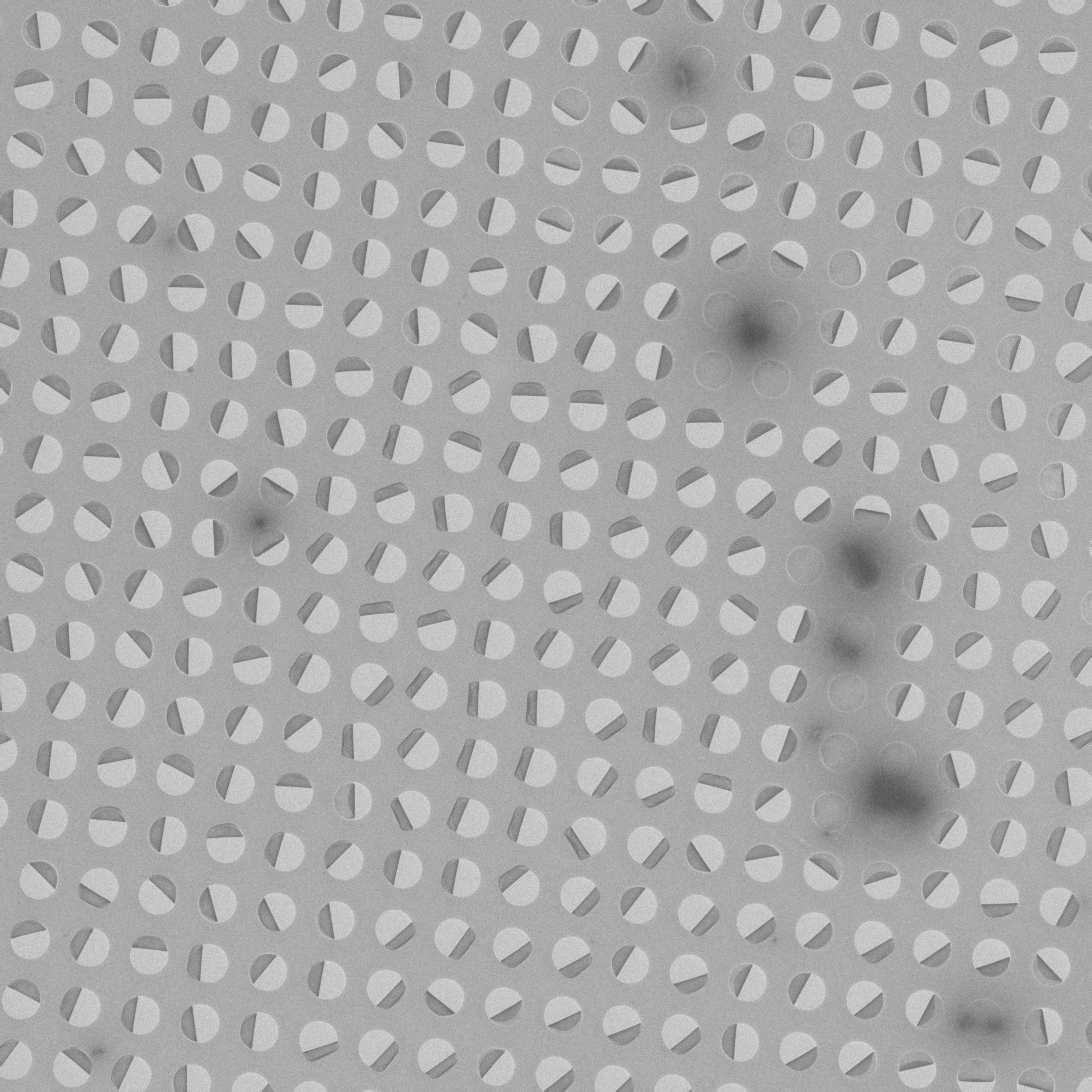
Submitted by: Mária Gondová, Pavel Plevka Research Group – Structural Virology, CEITEC MU
Title: Ugly 70's textile design
Description: The picture shows a holey carbon grid for electron microscopy that is covered with a 3 nm thick carbon film. However, the carbon film is broken in most of the holes like a lid of a can. Overall, it reminds me of the ugly (brownish) patterns on textiles popular in the 60's or 70's.
FEBRUARY

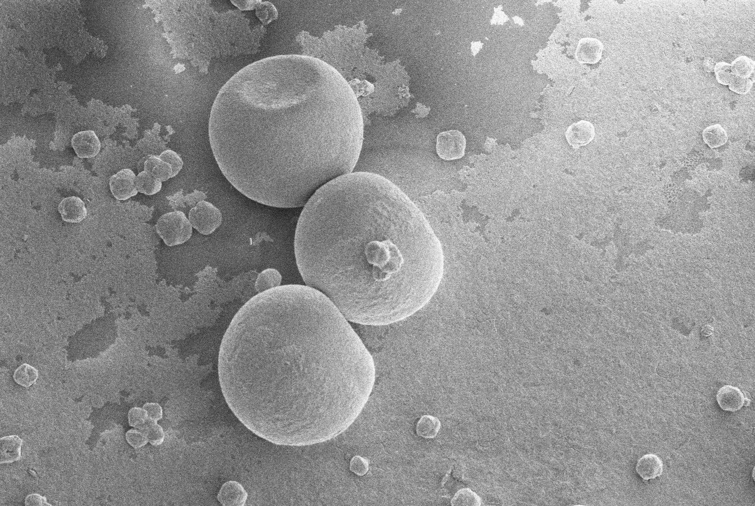
Submitted by: Michaela Procházková, Pavel Plevka Research Group – Structural Virology, CEITEC MU
Title: Microscopic snowflake
Description: Water crystals in the form of a flower or star appear regularly on cryoEM grids, and thus spoil the otherwise perfectly sound sample of Staphylococcus aureus cells. The side view of the crystal is captured by a gallium ion beam on the SEM Versa 3D instrument.
MARCH
Submitted by: Ondřej Jurček, Radek Marek Research Group – Structure of Biosystems and Molecular Materials, CEITEC MU
Title: Sorry (snow)man, spring is coming
Description: This SEM picture shows a beauty of self-assembly processes where small metallocomplexes, about 3.5 nm in diameter, derived from natural compounds, can self-organize into large spherical microparticles of about 85 μm in diameter. This concept can be used for development of novel porous metal-organic materials with a broad range of applications.
APRIL
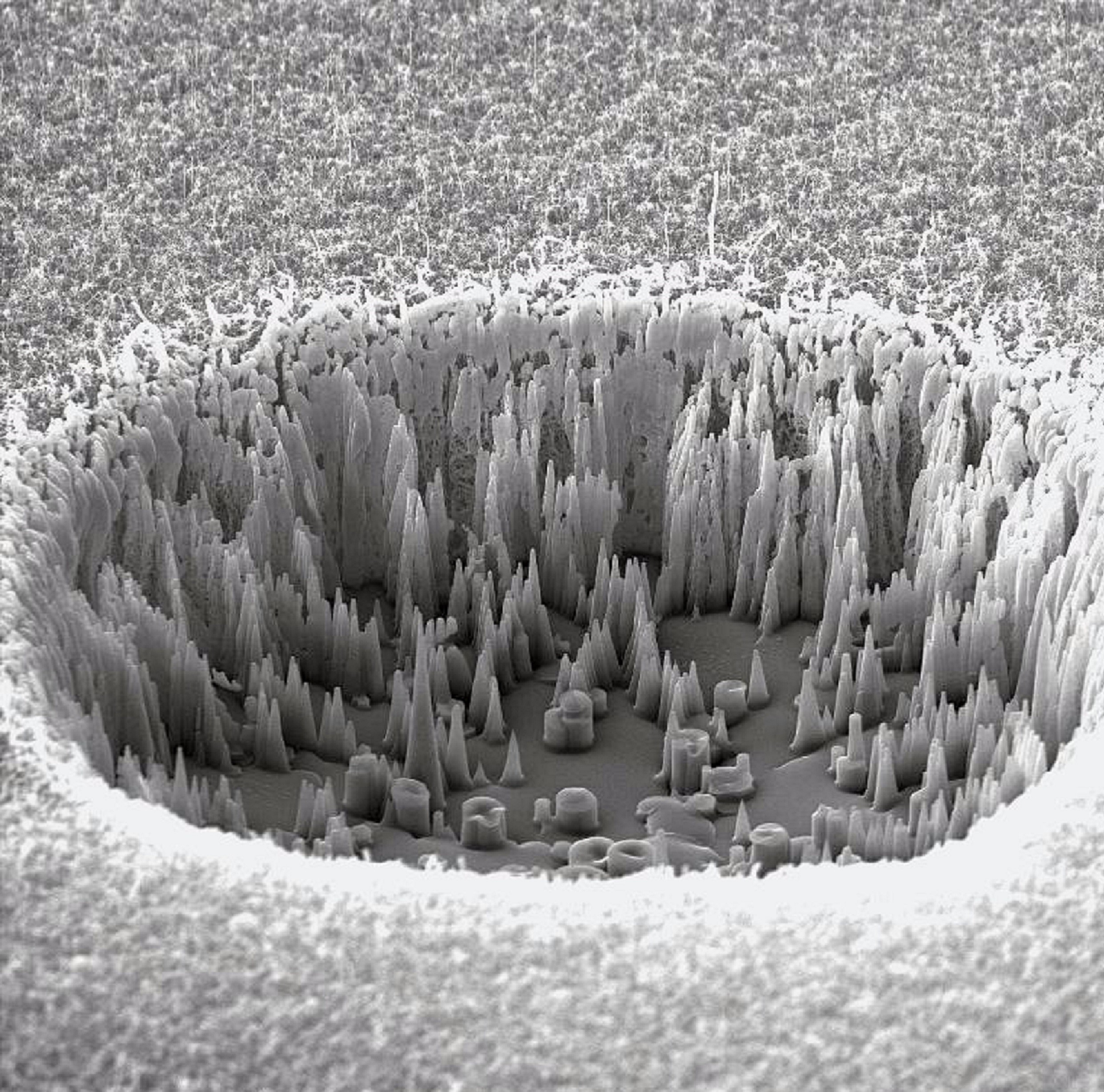
Submitted by: Preeti Kaushik, Lenka Zajíčková Research Group – Plasma Technologies, CEITEC MU
Title: CNT-Thorny nano well
Description: Carbon nanotubes (CNTs) fabricated on Si/SiO2 substrates using chemical vapor deposition (CVD). Characterized by focused ion beam milling- scanning electron microscopy (FIB-SEM) to evaluate their length.
MAY

Submitted by: Soňa Valuchová, Karel Říha Research Group – Plant Molecular Biology, CEITEC MU
Title: Ghosts of a failed experiment
Description: What is more scary than a failed experiment? We just love our ghost-like ovules of Arabidopsis thaliana imaged using light sheet microscopy.
JUNE

Submitted by: Sorin Tanasa, Karel Říha Research Group – Plant Molecular Biology, CEITEC MU
Title: Plants have feelings too
Description: Dedication to health workers on the COVID19 battleground.
JULY

Submitted by: Ján Bíňovský, Pavel Plevka Research Group – Structural Virology, CEITEC MU
Title: Viral Genome-based Death Star Design
Description: To reconstruct a 3-dimensional structure of a virus by cryo-EM in more detail, we can enhance a signal from the viral capsid by masking out a signal coming from a viral genome. You can see the result of a late-night failed mask generation trial combined with an early-afternoon coffee-boosted creative mood. For those untouched by the Force - the mask resembles the famous giant space station from the Star Wars movies, called the Death Star.
AUGUST

Submitted by: Marta Šiborová, Pavel Plevka Research Group – Structural Virology, CEITEC MU
Title: Smallest CEITEC Logo
Description: Even the hexagon is perfect and symmetrical. It is just ice contamination on my cryoEM grid.
SEPTEMBER

Submitted by: Subhasis Chattopadhyay, Radek Marek Research Group – Structure of Biosystems and Molecular Materials, CEITEC MU
Title: If you love your science, it will love you back
Description: Keep on doing hard work from your "heart." Even if you fail, they'll motivate you because "FAIL" is First Attempt In Learning. Picture courtesy of SEM-Versa3D.
OCTOBER

Submitted by: Lenka Šmerdová, Pavel Plevka Research Group – Structural Virology, CEITEC MU
Title: Olympic Rings
Description: When alignment doesn't go well - misaligned Z-stacks collected by Lightsheet microscopy.
NOVEMBER

Submitted by: Marek Večeřa, Ondřej Slabý Research Group - Molecular Oncology – Solid Cancer, CEITEC MU
Title: Artificial Intruder
Description: One day, I was taking photos of an adherent glioblastoma cell and found this random helix-shaped fragment. Luckily, it did not harbour any pathogens. There‘s no need for another epidemic!
DECEMBER

Submitted by: Thula Sravankumar, Tomasz Nodzynski Research Group – Mendel Centre for Plant Genomics and Proteomics, CEITEC MU
Title: Quarantined GFP tag protein inside the root cells
Description: Yes, it is GFP tag protein expression in Arabidopsis thaliana root cells, but it reminds me of self-isolation, always alone in a single cell, like nowadays, when people are quarantined during COVID-19. I hope we will get back our normal days soon.
Notice
At this time, the competition is on hold. Please stay tuned for any upcoming news. If you have any questions or technical related inquiries, please send them to Karolína Šupejová at karolina.supejova@ceitec.muni.cz.
Other Submitted Pictures
Photogallery


