
11. Apr. 2024
Marine algae such as the unicellular Emiliania huxleyi exert a cooling effect on the Earth's climate. However, the abundance and species diversity of algae in the ocean is affected by viral infections. The Structural Virology research team at CEITEC Masaryk University, led by Pavel Plevka, has now used cryo-electron microscopy to describe the structure and replication cycle of the virus EhV-201 infecting the alga Emiliania huxleyi. Their research shows how the alga defends itself against the infection and how the virus uses the resources of the host cell to produce its progeny.
Climate change is one of the most pressing societal challenges of our time and humanity will need to understand all its important contributing factors in order to mitigate them or at least adapt to the environmental changes and marine algae are one of the factors influencing the Earth's climate. Emiliania huxleyi belongs to a group of algae called coccolithophores because it produces tiny calcite disks, coccoliths, which coat its surface. The coccoliths reflect light like tiny mirrors, reducing the absorption of sunlight by the ocean. Coccoliths contain carbon and their deposition on the seabed leads to a reduction in the concentration of carbon dioxide in the atmosphere, limiting its contribution to the greenhouse effect. In addition, Emiliania huxleyi produces substances that trigger the formation of clouds and thus contribute to the cooling of the atmosphere. Emiliania huxleyi grows globally in oceans with tempered and cold waters, however, its population density is limited by coccolithoviruses such as Emiliania huxleyi virus 201 (EhV-201). Research by a group of structural virologists at CEITEC Masaryk University has now brought science a step closer to answering the question of how these viruses bypass the protective layers at the surface of Emiliania huxleyi cells and destroy these marine algae in large numbers.
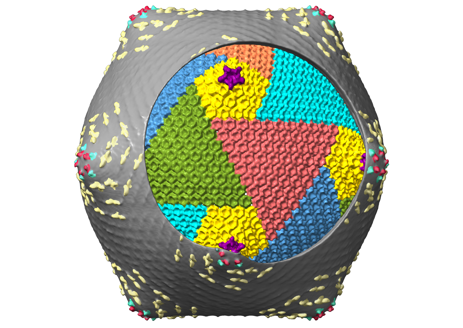
The researchers focused on EhV-201, a virus that commonly infects Emiliania huxleyi, but only little was known about its structure and replication cycle. Reconstructing the virus three-dimensional structure, however, was complicated by the discovery that EhV-201 particles are pleomorphic, meaning that each viral particle is unique. "We had to use a tomographic approach to extract the maximum information from each individual virus particle, and in the end, we showed that the virus particle is surrounded by two membrane layers, between which lies the virus capsid. The outer membrane of the virus is attached to the capsid by several types of transmembrane proteins." says Miroslav Homola, first author of the study. "EhV-201 particles infect Emiliania huxleyi by attaching to its surface and fusing the inner membrane of the virus with the plasma membrane of the cell, thereby delivering the virus genome into the cell." adds team leader Pavel Plevka.
In addition, by studying the virus life cycle directly in algal cells using unique approaches and a range of state-of-the-art visualisation and reconstruction methods, the CEITEC Masaryk University scientists were able to show that Emiliania huxleyi cells protect themselves from virus infection by several membrane layers, which have never been described in this alga before. "This armour enables Emiliania huxleyi cells to trick the virus into releasing its genome outside the cell, preventing infection. Some virus particles, however, find gaps in the protective envelopes, release their genome into the cytoplasm of the cell and initiate infection. Using modern combined electron and plasma microscopes, we cut thin lamellae of vitrified infected algae. We could thus observe how viruses form so-called virus replication factories at specific locations in the cell, where progeny virus particles assemble. The virus also removes the protective layers from the surface of the infected algae so that it can be continuously released into the environment and infect other cells" Homola concludes.
The scientists faced many challenges from the very beginning of the project, the results of which were published on 10 April 2024 in the journal Science Advances. CEITEC Masaryk University scientists work in a lab in the middle of Europe, but needed to simulate as closely as possible a natural environment in the ocean to successfully culture the algae. Transporting "live" seawater to a landlocked country was complicated and uneconomical, so they ended up using water from a local marine aquarium.
Shared laboratories at CEITEC Masaryk University equipped with state-of-the-art microscopes played an indispensable role in determining the structure of the virus. Moreover, the scientists could not use any standard reconstruction method, so they had to come up with a combination of unique approaches. As a result, they were able to describe in detail the structure of EhV-201 particle and its life cycle directly inside the cells of the alga Emiliania huxleyi.
Photogallery

Author

Expert
