
14. Jan. 2026
Researchers from CEITEC BUT participated in an international study that revealed the crucial role of specific genes in the development of the vocal and auditory apparatus in mammals. Using computed tomography, they created detailed 3D models of mouse embryos, which enabled them to describe the organ changes that occur after the inactivation of these genes. The results of the research, conducted in collaboration with French biologists, were published in the scientific journal PLOS One.
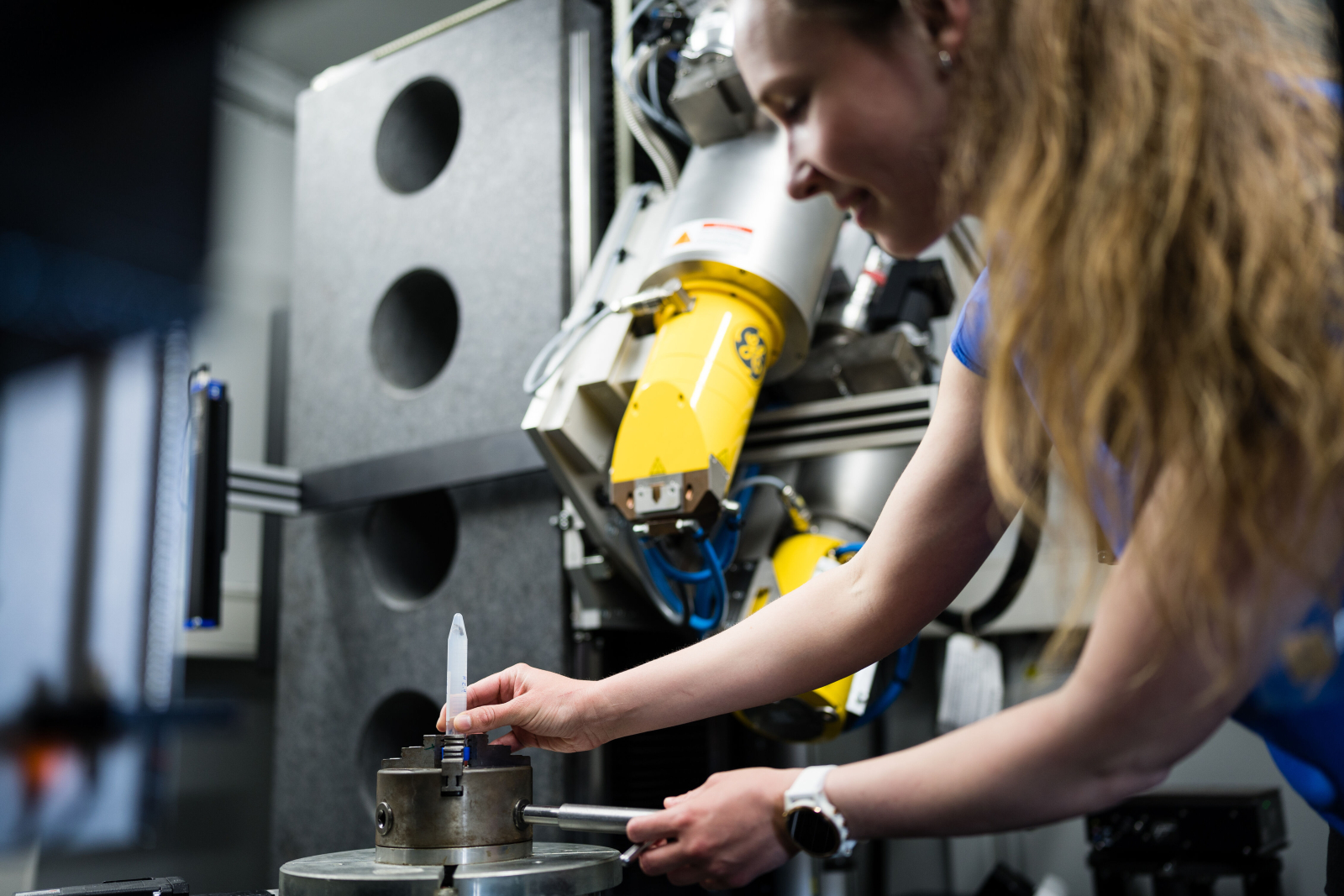
The Laboratory of X-ray micro- and nano-computed tomography (CTLAB CEITEC) at CEITEC BUT has long been involved in the development of non-destructive testing methods, which are widely used primarily in industry. However, this know-how is increasingly crossing the boundaries of technical fields and is also being applied in biology and medicine. An example of this is the collaboration with a French research team, in which scientists focused on the influence of selected genes on the development of the vocal tract and auditory system of mammals.
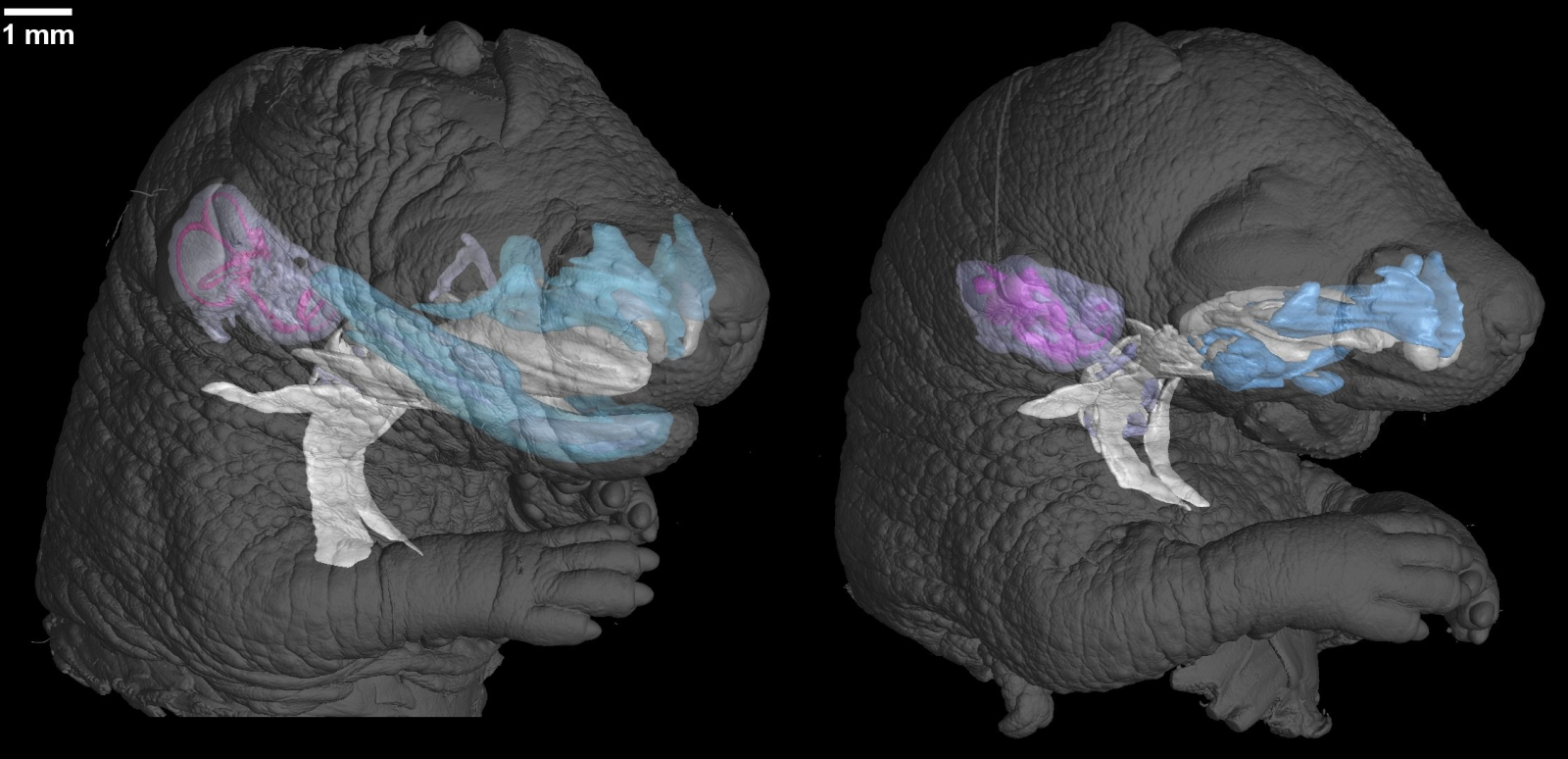
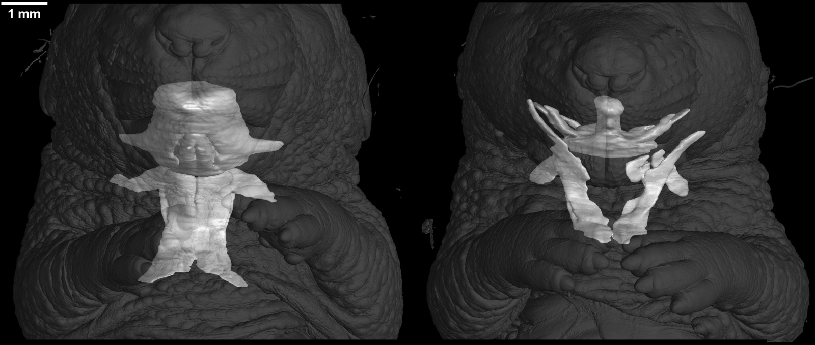
The task of the Brno scientists was to scan biological samples in detail and then create accurate 3D models. These allowed them to examine the internal structure of mouse embryos without damaging them and to accurately describe the changes resulting from genetic interventions.
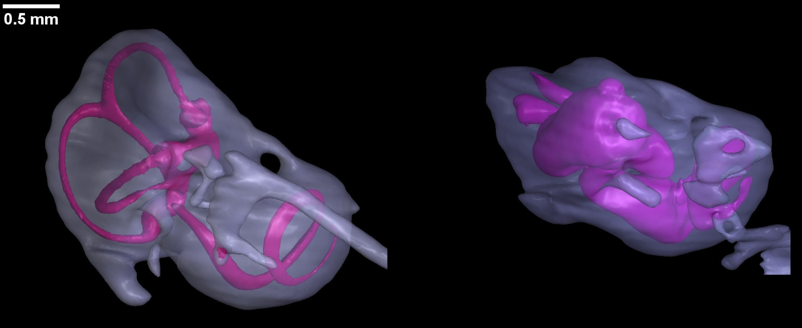
The study focused on the inactivation of Dlx5/6 genes in mouse embryos. These genes play a crucial role in the development of limbs, facial structures, and the vocal and auditory apparatus. The research was based on the concept of pleiotropy, which posits that a single gene can simultaneously influence multiple characteristics of an organism. It was this principle that led scientists to hypothesize that the Dlx5/6 genes may simultaneously shape all components of the mammalian acoustic communication system.
To accurately identify developmental changes, biologists combined classical histological methods with modern micro-CT tomography. This part of the research was undertaken by scientists from CEITEC BUT Markéta Kaiser and Tomáš Zikmund. Using computed tomography, they created detailed three-dimensional models of embryos, which allowed them to compare healthy and genetically modified samples.
Tens of thousands of images and hundreds of hours of precise work
Computed micro-tomography works on the same principle as medical CT, but with significantly higher resolution – up to five micrometers, which is the size of a red blood cell. “Unlike medical devices, where the radiation source and detector rotate around the patient and the time is carefully monitored to ensure that the examination is safe for humans, we did not have to worry about this because the samples were not alive. Therefore, when scanning each of them, we only rotated the test tube, which is a procedure that guarantees higher resolution due to higher radiation doses,” explains Kaiser.
Each sample was recorded in approximately 2,000 images over several hours of scanning, gradually creating an extensive data file. Because X-rays naturally have difficulty imaging soft tissues, it was necessary to stain the samples with heavy metal solutions before scanning.
The most demanding part of the work came after the scanning itself. The scientists had to analyze tens of thousands of grayscale image slices and, in collaboration with French biologists, accurately identify individual organs and tissues. The subsequent reconstruction of 3D models required repeated checks and detailed comparisons with the original data. “We were among the first to start using and systematically developing this method at CEITEC. Thanks to it, we can visualize even very fine structures, such as cartilage, and observe their spatial arrangement,” adds Kaiser. Part of the process is now accelerated by artificial intelligence tools that help automate the recognition of specific structures – similar to how AI is used, for example, in the analysis of medical CT images.
The results of the study showed that inactivation of the Dlx5/6 genes causes severe disorders of the musculoskeletal system of all organs of the vocal tract, while also affecting the development of the outer, middle, and inner ear. The researchers thus clearly demonstrated the pleiotropic role of these genes in the formation of the complete vocal and auditory apparatus of mammals.
CEITEC technologies open up new possibilities for biological research
According to Markéta Kaiser, the successful collaboration with the French team was made possible by the laboratory's long-term experience with the application of computed tomography in biological research. In the past, scientists from CEITEC BUT have participated in several projects focused on the development of organs and tissues that require a detailed yet non-destructive view of the internal structure of samples.
“This project clearly shows how cutting-edge imaging technology can help biologists answer questions that would be very difficult or impossible to address using other methods. Thanks to 3D models, we were able to accurately describe how the inactivation of Dlx5/6 genes affects the entire complex of organs responsible for voice and hearing. It is precisely the combination of technical and biological approaches that is the key to understanding such complex developmental processes,” concludes Markéta Kaiser.
Author: Kristina Blűmelová
Photogallery
Expert
