We provide X-ray submicron computed tomography (Submicron CT) for non-destructive three-dimensional visualization and inspection of a wide range of samples at exceptionally high resolution. The method delivers detailed information about the internal structure of materials and biological specimens while preserving their native state.
Method
Submicron CT enables imaging of samples at cellular resolution in a laboratory environment without damaging the specimen. The simple sample preparation preserves native structures and facilitates reliable interpretation of the results. The technology provides comprehensive information across multiple length scales: from macroscale structures to submicron details and supports both qualitative visualization and quantitative analysis.
Typical applications include:
- Phenotype evaluation
- Structural morphology analysis
- Tissue and cellular feature analysis
- Root system analysis
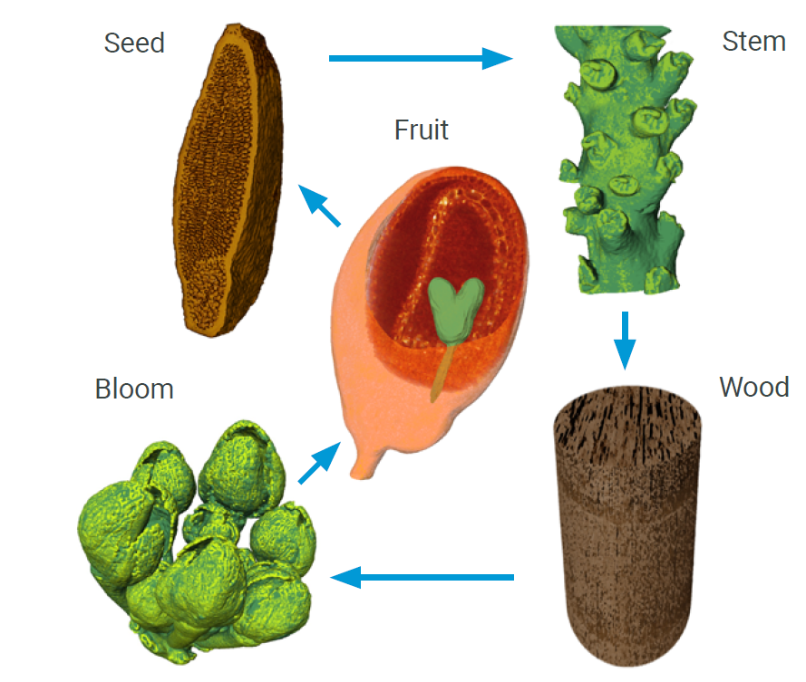
- Seed, fruit, and embryo analysis
- Vascularization analysis
Equipment
The analyses are performed using the Rigaku nano3DX X-ray microscope (XRM), designed for non-destructive imaging of relatively large samples at very high spatial resolution. The system combines a high-powered rotating anode X-ray source with a high-resolution sCMOS detector, enabling rapid data acquisition and optimized imaging conditions for a wide variety of sample types.