 Share
Share
About Event
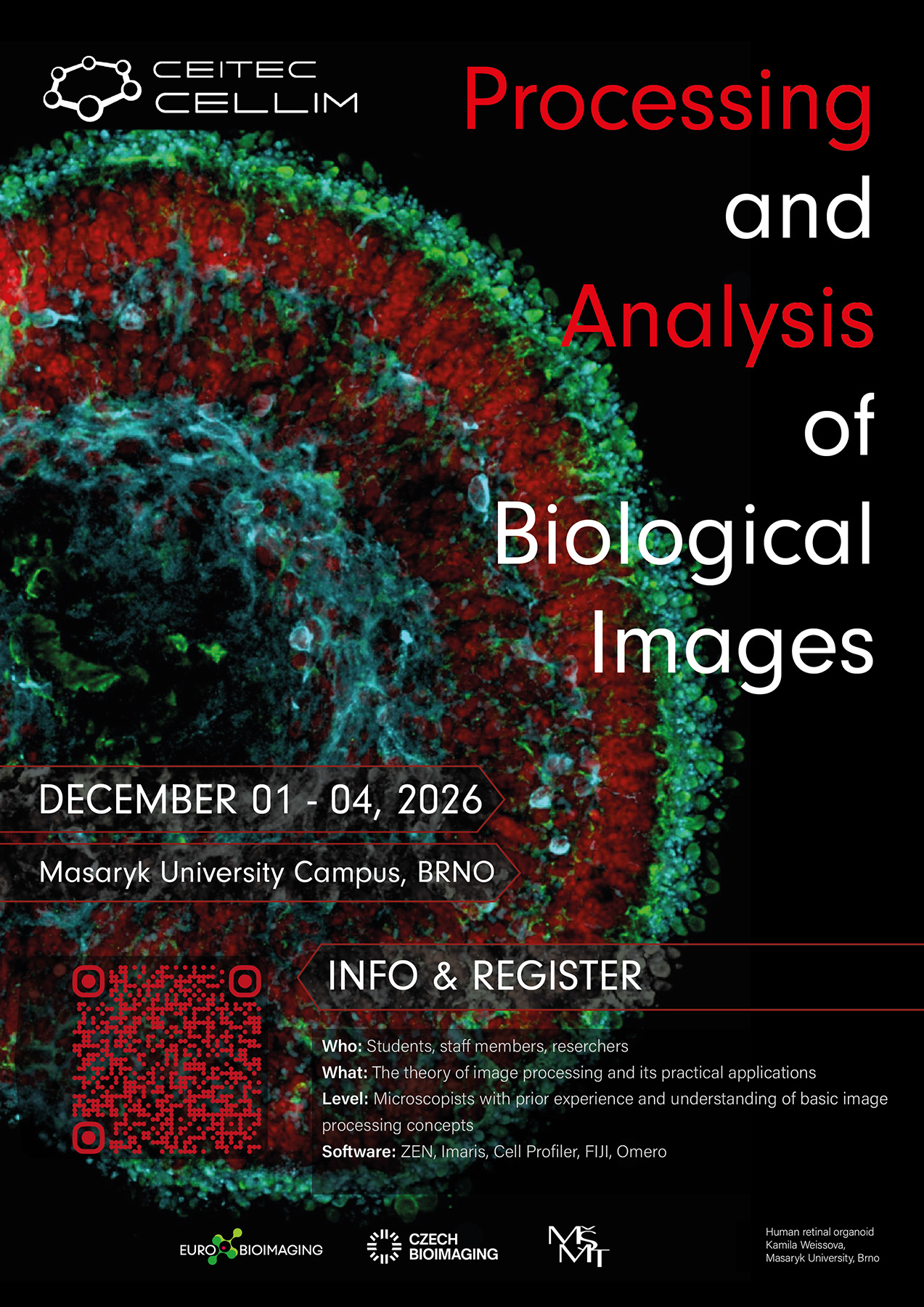
Modern microscopy generates increasingly complex and information-rich datasets, making appropriate image processing, visualisation, and quantitative analysis essential parts of biological research. This four-day course is designed for biologists who want to develop a practical understanding of biological image processing and gain hands-on experience with commonly used image analysis platforms.
The course combines essential theoretical concepts with practical exercises covering the complete image-analysis workflow, from digital image formation and preprocessing to segmentation, object detection, tracking, quantitative analysis, visualisation, and data management.
Participants will work with both open-source and commercial software, including Fiji/ImageJ, SAMJ, ZEISS ZEN, Imaris, and OMERO. Particular attention will be given to selecting appropriate analysis approaches, understanding how processing steps influence quantitative results, and developing reproducible workflows for microscopy data.
The course also introduces modern approaches to bioimage data organisation and sharing, including FAIR data principles, image repositories, OME-Zarr, and the OMERO data-management environment.
The course is organised by the Cellular Imaging Core Facility, CEITEC Masaryk University, in collaboration with the Faculty of Informatics, Masaryk University.
Course content
Day 1 – Fundamentals of digital images and open-source image analysis
Introduction to microscopy detectors and digital image formation, followed by the fundamental principles of image processing and quantitative image analysis. Participants will become familiar with Fiji/ImageJ and explore segmentation workflows using the SAMJ plugin. The day will also introduce bioimage repositories, FAIR data principles, and modern microscopy data formats including OME-Zarr.
Day 2 – Image processing and analysis in ZEISS ZEN
Practical introduction to working with microscopy datasets in ZEISS ZEN beyond image acquisition. Participants will explore image preprocessing, visualisation, measurements, segmentation, and the analysis tools available within the ZEN Analysis environment.
Day 3 – 3D visualisation and quantitative analysis in Imaris
Hands-on work with Imaris focusing on multidimensional microscopy datasets. Topics will include 3D and 4D visualisation, segmentation of biological structures, object detection, tracking, quantitative measurements, and extraction of biologically meaningful information from image datasets.
Day 4 – Image data management with OMERO
Introduction to the OMERO repository environment and good practices for managing microscopy datasets. Participants will learn how to organise, annotate, access, visualise, and share imaging data and how repository-based workflows can support FAIR and reproducible research.
Learning outcomes
After completing the course, participants should be able to:
- understand the fundamentals of digital image formation and image processing;
- perform basic image processing, segmentation, and quantitative analysis;
- use Fiji/ImageJ, ZEISS ZEN, and Imaris for biological image analysis and visualisation;
- understand FAIR data principles and manage microscopy datasets using OMERO and modern bioimage data formats.
More information
LEVEL/PREREQUISITE: Microscopists with prior experience and understanding of basic image processing concepts (Recommended previous level: Fundamentals of Light Microscopy)
COSTS (including coffee breaks and refreshments):
Academic: 3, 500 CZK + VAT,
Others: 8, 500 CZK + VAT
INFO & CONTACT: