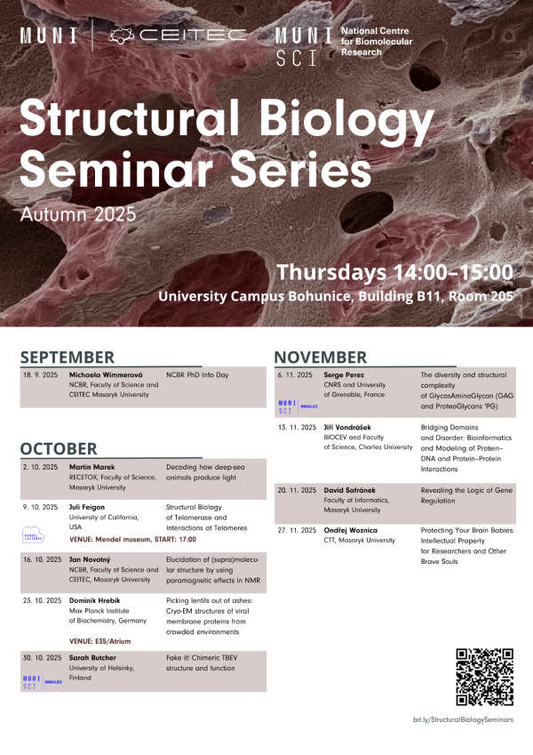
About event
Cryo-electron microscopy single-particle analysis traditionally requires particles of high purity embedded in thin sections of vitreous ice. It has been thought that characterisation of more complex samples, such as heterogeneous enveloped viruses, necessitates cryo-electron tomography, which separates densities in the third dimension. However, data collection and processing of electron tomograms are slower than those for single-particle analysis. Recent advances in hardware and software now allow a wider range of structures to be resolved in crowded environments using single-particle analysis. By employing machine-learning-based picking of objects of interest and subsequent deep classifications, I determined in situ structures of individual proteins at up to 2.1 Å resolution from HIV-1 particles with an average thickness of approximately 120 nm. I demonstrate that it is possible to resolve the structure of the peripheral HIV-1 membrane protein, the matrix protein, with a molecular weight of less than 500 kDa, to sub-3 Å resolution. These structures have resolved a long-standing mystery in HIV-1 biology – the HIV-1 matrix protein binds a small conserved spacer peptide 2, with a previously unknown function. The binding triggers rearrangements of the matrix protein and viral membrane that regulate HIV-1 fusion with the host cell. Additionally, I will present potential future developments and applications of the approach, especially in the context of high-throughput in situ structural biology of membrane proteins and in-cell structural proteomics.
The lecture is part of the Structural Biology Seminar Series. PhD candidates, postdocs, and research group leaders from the structural biology field present their newest research results to the MUNI scientific community. Ideal space for knowledge sharing, networking and professional growth.


 Share
Share
