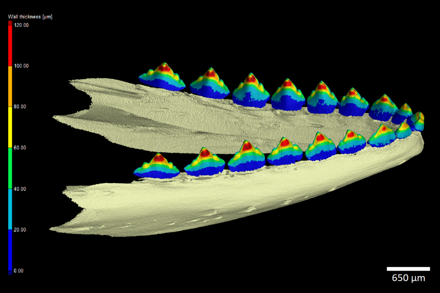
Micro-CT data enables the creation of accurate 3D models of individual organs and complex structures. Professional software tools allow for a wide range of analyses, including measurements of lengths, volumes, and wall thicknesses, as well as shape comparisons. Combined with histological methods, these analyses provide comprehensive characterization of the samples under investigation. Our activities focus on adapting specialized industrial tools for biological applications and introducing new methods of data interpretation, such as 3D printing (Tesařová et al., Journal of Instrumentation, 2016) and interactive 3D PDFs (Tesařová et al., Scientific Reports, 2019).
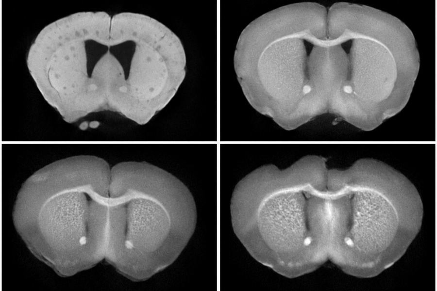
Micro-CT scanning of samples in their natural state primarily provides information about hard tissues, such as teeth and bones, due to their ability to absorb X-rays. To visualize soft tissues, contrast-enhanced micro-CT imaging is required. Contrast enhancement is achieved using solutions containing elements with high atomic numbers, such as iodine, or compounds such as phosphotungstic acid (Zikmund et al., Journal of Instrumentation, 2018). However, contrast staining protocols must be optimized for each specific type of organ or tissue. Our research therefore focuses primarily on developing and optimizing new staining protocols for the visualization of cartilage (Kaucka et al., eLife, 2018), liver, brain, and heart tissues.
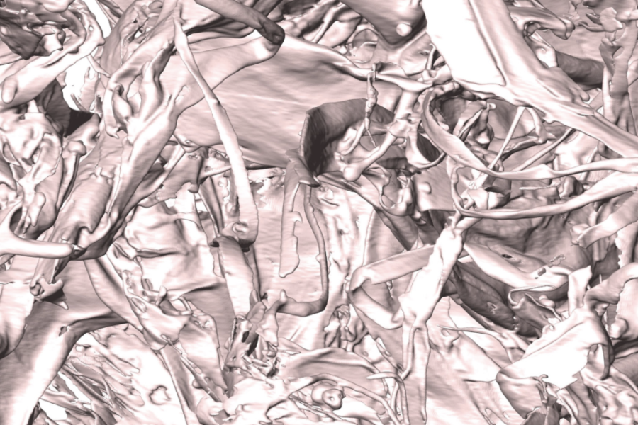
Tissue engineering focuses on the development of biocompatible materials designed to support, regenerate, or replace biological tissues. Newly developed materials can promote cell growth and facilitate the regeneration of damaged or diseased tissues. X-ray computed tomography (CT) is a powerful tool for the precise characterization of these complex three-dimensional structures, including cryogels (Sedlačík, Soft Matter, 2017), hydrogels, hydroxyapatite-based materials (Šťastný, J. Am. Ceram. Soc., 2018), and polymer scaffolds (Vojtová, J. Microsc., 2018). Our research focuses on developing methods for imaging scaffolds at submicrometer resolution and quantitatively characterizing their internal structures (Kalasová, Microsc. Microanal., 2018). The results of CT analyses are subsequently validated using 2D imaging techniques, particularly scanning electron microscopy (SEM).