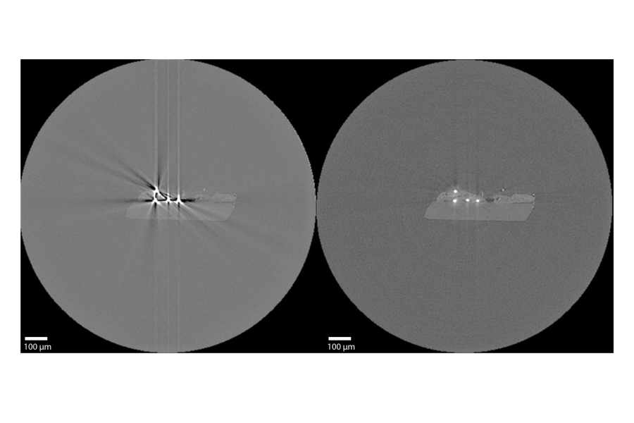
Phase Contrast Imaging (PCI) extends the capabilities of CT for analyzing materials with low absorption contrast, such as polymer structures or soft tissues. When X-rays pass through these materials, they cause only minor changes in intensity but also induce shifts in the phase of the radiation. Specialized PCI techniques make it possible to convert these phase shifts into image information, revealing structures that are difficult to distinguish using conventional CT. Although PCI has traditionally been used primarily at synchrotron facilities, advances in X-ray sources and detectors are enabling its increasingly widespread application in laboratory CT systems. Our research focuses on determining the requirements that CT systems must meet to enable propagation-based phase contrast imaging (Kalasová, IEEE TIM, 2019) and developing methodologies for the practical implementation and application of PCI.
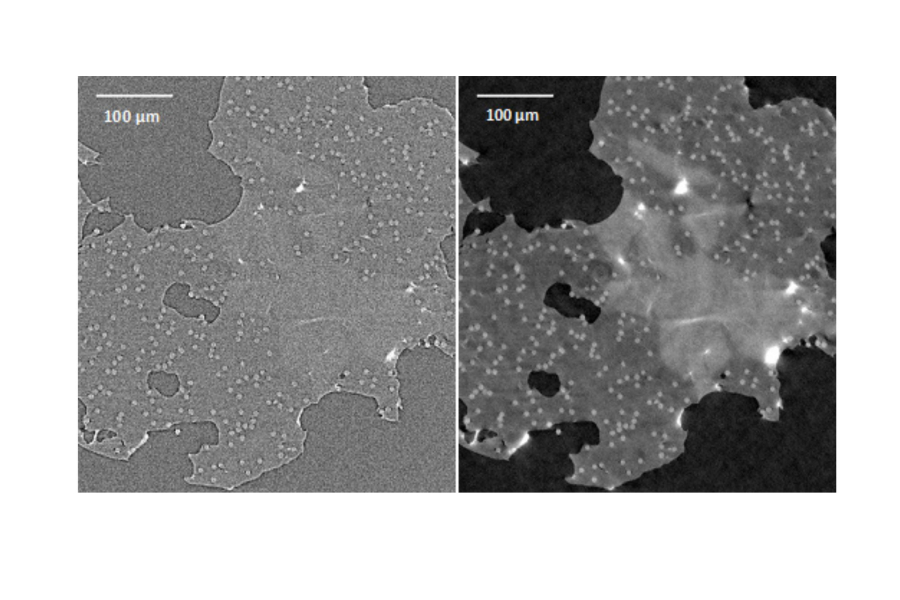
Dual-Energy Computed Tomography (DECT) uses two distinct X-ray energy spectra to distinguish individual components of a sample—materials or tissues—based on differences in their X-ray attenuation properties. This approach makes it possible to differentiate materials that are difficult or even impossible to distinguish using conventional CT. The main advantage of DECT is therefore more accurate material segmentation and classification. Our laboratory focuses on implementing DECT in laboratory nano-CT systems and developing a comprehensive methodology for data acquisition and processing. Our research aims to maximize the potential of this technology, particularly in reducing metal artifacts and developing new methods for segmenting biological soft tissues.