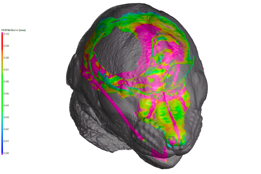
Computed tomography (CT) is a key tool in biology, enabling non-destructive 3D visualization and analysis of biological structures. Detailed imaging of bones and soft tissues allows for the creation of accurate 3D models and the study of anatomical features and developmental processes, including ossification. CT is also used in the development of bone substitutes, implant evaluation, and characterization of soft tissues such as muscles, organs, and blood vessels. It also enables the investigation of foreign particles in animal bodies, such as microplastics, providing valuable data on the impact of environmental factors on health. By offering detailed insights into internal biological structures, CT contributes to advances in developmental biology, regenerative medicine, and environmental biology, fostering a deeper understanding of biological processes.
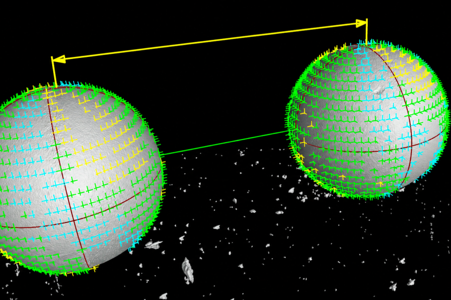
Computed tomography (CT) is an important tool in modern metrology, enabling precise and non-destructive measurement of both external and internal structures of objects. Research focuses on evaluating spatial resolution, determining measurement uncertainties, and developing reference objects and methods for verifying the accuracy of CT systems. The aim is to improve the reliability, accuracy, and metrological traceability of CT measurements for both research and industrial applications.
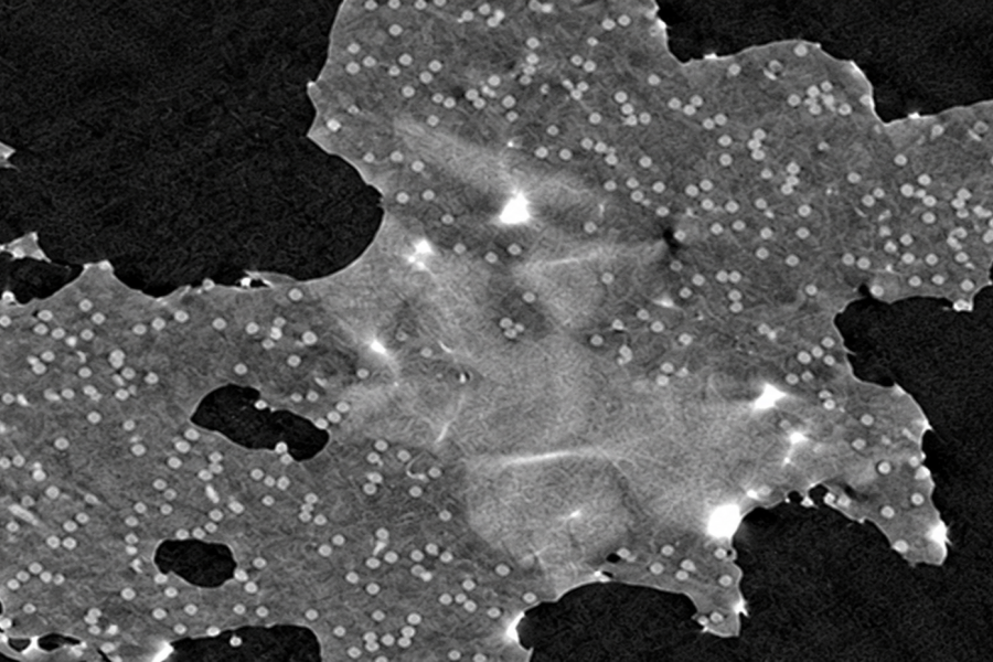
X-ray computed tomography (CT) technology is continuously evolving. New imaging techniques are being developed and implemented to provide more comprehensive information about internal structures.
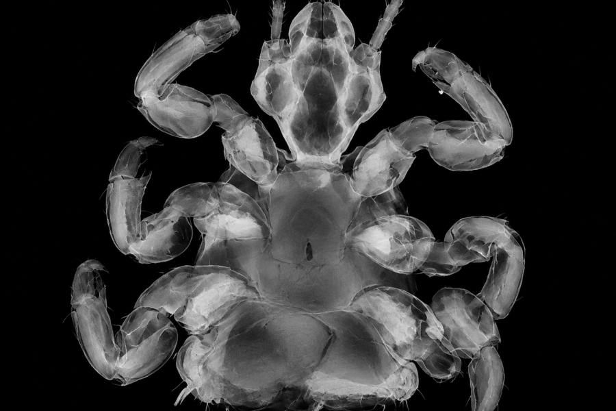
Computed tomography (CT) relies heavily on the performance of computer hardware combined with specialized data processing and reconstruction software. While numerous general-purpose software packages for tomography are available, certain samples require highly specific approaches to data processing and analysis that are often not commercially available. At CT Lab, we focus on developing custom software solutions to improve the quality of tomographic data and facilitate its analysis, ranging from artifact reduction to efficient automated post-processing.
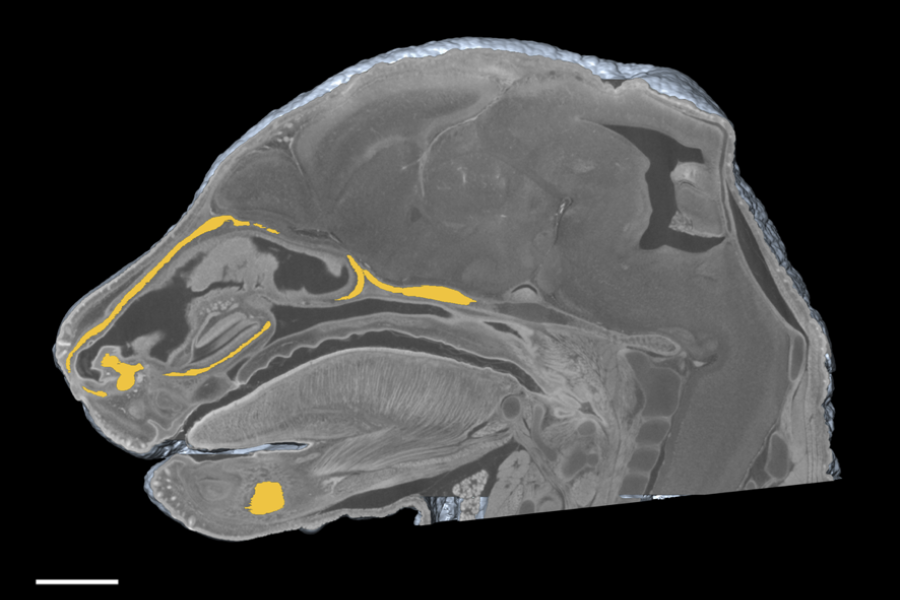
Extracting relevant information from acquired CT data is one of the most important stages of the entire CT analysis process. Each CT measurement presents new challenges that, in some cases, exceed the capabilities of available software tools. To address these specific challenges, we develop custom software solutions based on the latest computer vision techniques, including artificial intelligence (AI).
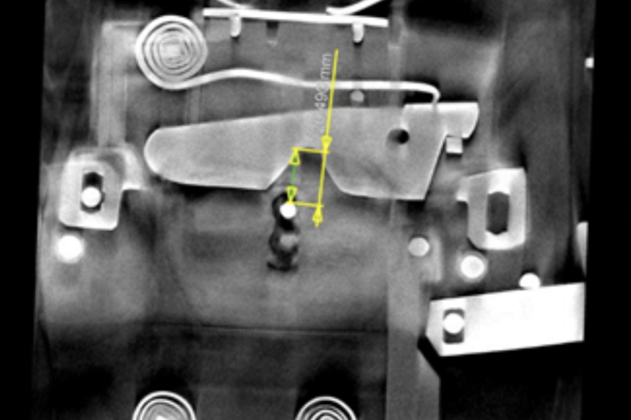
Our laboratory uses advanced computed tomography (CT) techniques to non-destructively investigate the internal structures of materials, natural specimens, and valuable historical objects. Through detailed 2D and 3D imaging, we can explore beneath the surface of rocks, minerals, fossils, archaeological artifacts, and works of art without causing damage. CT data enables us to reveal hidden structures, cracks, and cavities, assess porosity, density, and the distribution of different materials, and investigate the condition and history of the objects under examination.By combining CT with other analytical methods, we gain a comprehensive understanding of each sample, from its internal structure to its material properties and chemical composition.
A demonstration of our biological research – analysis of a mouse skeleton and brain. The analyses were performed using VGSTUDIO software.

